2D radiographic scans have become common practice to explain complex injuries in personal injury work practices. If it’s good enough for doctors then surely it’s good enough for lawyers, right?
Here’s the thing, courtroom visuals have evolved over the last number of years.
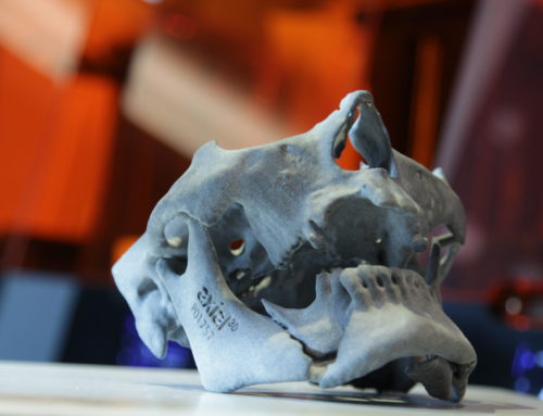
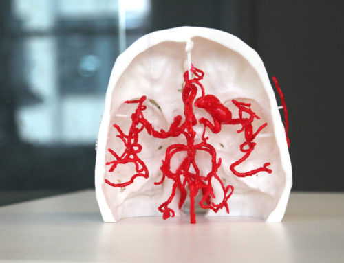
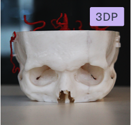
We now have access to medical illustrations, video re-enactments, and now… 3D printed models created from your client’s medical scans that can show their exact injury.
Lawyers across the country now have access to these incredibly lifelike replicas of their client’s anatomy, a medical-grade visualization designed to be understood by doctors and non-medical professionals alike. Each exhibit is unique to the client and their injury, bringing a new dimension to the traditional 2D radiographs we have become reliant upon.
So what is the difference between these 3D exhibits and 2D radiographs or medical illustrations?
Effective visuals resonate in a juror’s mind long after their presentation during the trial or mediation. Critically, if the juror doesn’t understand what he or she is looking at, it just won’t make that lasting impression. While traditional visuals such as 2D radiographs may convey what is required, a 3D replica enables the juror to navigate and examine the exhibit, which helps them to understand the problem and build empathy with your client.
This free infographic helps to break down the differences between the types of imaging available for use in court right now.

Read more: Find out how 3D exhibits are transforming juror’s understanding of injuries