A step beyond 2D imaging

3D printing is transforming personal injury cases.

Using 3D visuals to demonstrate long-term effects of personal injury cases

Danielle Blandford is an attorney at Morgan & Morgan, the largest personal injury firm in America. She specializes in automobile cases – helping people who have been injured in car crashes caused by negligence.

In a recent case, her client had been involved in a motorcycle accident. He was impacted by a semi-truck in 2017 and landed on his right hip. When looking at the X-ray from the day of the accident, no real fracture or internal damage could be seen. However, four years later, the hip was in complete disarray and the client needed a hip replacement.

Ms. Blandford’s client had an underlying condition of arthritis, which is where the difficulty of this case arises. She had to prove to the jury that this hip replacement was needed because of the accident, which caused damage to the external portion of the hip but did not actually cause any type of fracture.

Legal3D provided two 3D visuals for this case: one made based on the X-rays of the day of the accident and another one based on the X-rays of one year after the accident. The medical expert explained that the difference in the condition of the hip between those two images looked like they were ten years apart, instead of just one, demonstrating the extent that the trauma has worsened the client’s arthritis. 

“I would recommend Axial3D because you need to be able to show the injury and have people understand why it’s painful. And the only way you’re gonna be able to do that is if you have a model or you have an image that you can show them to compare a normal body part versus your client’s body part, where it was injured. And that’s where people understand it.”


Thanks to the help of the 3D visuals, the jury was able to understand the long-term effects of the trauma. As a result, they decided in favor of Ms. Blandford’s client. “I think as an attorney, your job is not only to tell the jury that your client is in pain […]. I believe your job as an attorney is to explain it. I feel like that’s my job and Legal3D is the way that I do it at trial.”

2021-11-26T11:01:04+00:00November 26th, 2021|

Can innovation in healthcare imaging transform personal injury litigation?

You’ll know that any legitimate claim made in court must be supported by strong evidence. When it comes to personal injury claims, medical evidence is one of the most useful types of evidence – if not the most. Medical images can help solve many cases as they show everyone involved in the case how and where an injury was sustained. Afterall, they don’t say that a picture is worth a thousand words for nothing! Discover more about how our exhibits can improve jurors understanding.

If a picture is worth a thousand words, what is the value of a 3D model?

Medical images such as 2D scans or illustrations can often be very complex and difficult to understand, even for medical professionals – never mind jurors! The use of 3D exhibits can help overcome this problem and transform PI litigation. This transformation includes supporting faster settlement decisions, by helping mediators understand injuries more clearly. 

In fact, a recent study from Cranfield Forensic Institute showed that Jurors understanding of technical language used in the courtroom had increased to 94% with 3D exhibits compared to 79% with photographic evidence such as MRI and CT scans. Medical imaging can be so powerful when it comes to providing evidence in courts, and medical-grade 3D exhibits really do go a step further in helping jurors make more informed decisions, resulting in a more fair trial and a better chance at a faster, higher settlement.

We have been doing our bit to help ease the effect caused by COVID-19!

Our 3D exhibits are here to help with the current case backlog due to the COVID-19 pandemic and this is made possible as the use of these 3D models will ensure that decisions made by jurors in court are made a lot quicker because their understanding of the plaintiffs injuries are a lot better leading to quicker and better settlements.

Be an early adopter and help transform how personal Injury litigation cases are handled! Use Legal3D to help you with your personal injury cases and you won’t regret it.

2021-05-24T09:55:08+00:00February 24th, 2021|

Adapting Personal Injury work practices during the COVID-19 pandemic

The COVID-19 pandemic has had a huge impact on most sectors around the world, both economically and because firms have needed to adapt and reorganize. This includes those in legal personal injury.

Due to many courts being closed for the past 12 months, new ways of working have had to be implemented to allow law firms to continue their business operations

This includes communication and engagement with plaintiffs, defence teams, expert witnesses, and the courts remotely. Additionally, courts are not planned to open until 2022 in some states, making it increasingly more difficult to get personal injury cases resolved quickly. Due to these remote mediations taking place online, attorneys need the most efficient way to solve these cases.

This is where 3D virtual exhibits come in! Not only are they 100% accurate, but they can be produced and ready for use within 24 hours and most importantly they transform the understanding of all parties involved as medical professionals can explain the injuries using these exhibits far easier than a 2D medical illustration. The traditional method of using Medical illustrations are hard for non-medical professionals to understand, especially when typical personal injury work practices are being done remotely, but they can also be very time consuming to produce. There is also the option of getting numerous 3D exhibits printed and delivered to all parties for an even clearer understanding of the injuries or medical conditions.

Contact us here to have a dedicated success manager that will hep you with whatever you need.

Furthermore, if you would like to read more on how healthcare imaging will help transform personal injury litigation visit here.

2021-05-21T12:25:36+00:00February 24th, 2021|

Can a client-specific 3D exhibit enhance a juror’s understanding of complex injuries?

Today, jurors are being asked to make decisions based on the information gleaned from the same complex 2D medical images that doctors have spent years training to interpret. Without such adequate  training, there is a risk that the full severity of the injury may not be conveyed, or worse, the information could be misinterpreted by the untrained jurors. This doesn’t stop the jurors from making informed decisions, but it does make the process much more difficult than it needs to be – for the jury, the prosecution and everyone involved. 

So how can you as an attorney support jurors to better understand the severity of your client’s injuries? 

Our jury system began in the early 1600’s, and as time has moved on, so too has our access to technology and its benefits. CT and MRI scanning has been around since the 1970’s, and when first introduced, revolutionized how we see inside the human body. Lawyers and doctor witnesses continue to use these 2D scans to explain injuries to juries.

So how has this changed over the years?

Recent advancements in medical imaging have once again revolutionized how we visualize injuries. One example of such innovation is that of medical 3D modeling. Surgeons across the US are using our medical 3D printed models of their patients’ anatomy derived from the 2D medical scans to get a more detailed, realistic view of the injury or deformity they are treating.  The same medical-grade 3D models are now being used in court.

The ability to present a client’s exact complex injuries using 3D exhibits created from their own medical scans has transformed personal injury cases across the US. The 3D models alleviate the uncertainty experienced by the jury with complex 2D medical scans while acting as a tactile visual reference point for all members as they make decisions in deliberation.

 To see the difference between 3D exhibits and these other methods such as 2D scans and illustrations, visit our website here.

Read more: To see how 3D exhibits can help transform remote personal injury arbitration and mediation cases during COVID-19


2021-05-21T10:30:42+00:00February 24th, 2021|

Courtroom visuals: The difference between 2D and 3D exhibits

2D radiographic scans have become common practice to explain complex injuries in personal injury work practices. If it’s good enough for doctors then surely it’s good enough for lawyers, right?

Here’s the thing, courtroom visuals have evolved over the last number of years.

We now have access to medical illustrations, video re-enactments, and now… 3D printed models created from your client’s medical scans that can show their exact injury. 

Lawyers across the country now have access to these incredibly lifelike replicas of their client’s anatomy, a medical-grade visualization designed to be understood by doctors and non-medical professionals alike. Each exhibit is unique to the client and their injury, bringing a new dimension to the traditional 2D radiographs we have become reliant upon.

So what is the difference between these 3D exhibits and 2D radiographs or medical illustrations?

Effective visuals resonate in a juror’s mind long after their presentation during the trial or mediation. Critically, if the juror doesn’t understand what he or she is looking at, it just won’t make that lasting impression. While traditional visuals such as 2D radiographs may convey what is required, a 3D replica enables the juror to navigate and examine the exhibit, which helps them to understand the problem and build empathy with your client. 

This free infographic helps to break down the differences between the types of imaging available for use in court right now. 

Read more: Find out how 3D exhibits are transforming juror’s understanding of injuries

2021-05-21T12:27:30+00:00February 24th, 2021|

How lifelike 3D exhibits helped settle a $3.5 million personal injury case

When a Lancaster woman was hit by a truck in 2016, the top neurosurgeon at Gates Vascular Institute was called to treat serious injuries to her head and neck. Dr. Elad Levy was confident he could perform the procedures that would help her overcome the injuries. But he wasn’t sure he could lay out the steps he took when it came time to explain them to a jury.

“The anatomy and the injuries were so complex that I didn’t feel I could adequately explain them,” he said.

It turned out he didn’t have to. A Buffalo personal injury law firm landed a $3.5 million legal settlement on the eve of the trial last month using a new form of evidence created on the Buffalo Niagara Medical Campus: colorized 3D models made with the plaintiff’s black- and-white imaging scans that showed damage caused in the crash.

“Lawyers have spent years trying to explain these types of injuries to jurors while a doctor is pointing to different shades of gray on an MRI or X-ray film 20 feet from the jury,” said attorney John J. Fromen Jr., who represented the injured woman.

“Some jurors get it. Some don’t. For jurors to hold a model that shows the injuries, take it with them to deliberate, is a real game changer.” Lawyers and doctors involved with the civil lawsuit believe it may be the first time personal, anatomically correct, three-dimensional medical models were used in a U.S. personal injury case.

Fromen and his partner, Frederick G. Attea, said it won’t be the last. They predict attorneys who bring these kinds of cases, and defend lawsuits, will clamor for such persuasive evidence as 3D modeling improves and its costs come down.
Meanwhile, advances on the Medical Campus that put the University at Buffalo and Gates Vascular Institute along the forefront of neurosurgery and medical technology allowed doctors and lawyers to help the injured victim address and compensate for her injuries.

The crash

On a midmorning in March 2016, a commercial truck driver traveling about 55 mph on a rain-slicked, 40 mph zone in Lancaster lost control of his vehicle, which skidded over a curb and grass median, and struck a part-time bookkeeper as she retrieved the mail for a family business.
The passenger side mirror of the truck slammed into the woman’s head. A metal running board below the passenger side door tore open her right leg. Fromen and Attea described the crash and aftermath on condition that the woman, truck driver and company not be named, a condition of the legal settlement.
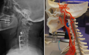
The woman, now 58, spent a week in intensive care at Erie County Medical Center, and 23 days in the hospital after the crash, as doctors stabilized her injuries and repaired fractures in her leg. Scans conducted on the woman’s head and neck also showed aneurysms behind both her eyes and damage to several discs in her cervical spine.
Levy – chair of the Department of Neurosurgery in the University at Buffalo Jacobs School of Medicine and Biomedical Sciences, and director of neuroendovascular services at Gates Vascular Institute – was assigned to deal with the head and neck injuries because of their complexity. In the following months, he used a catheter to insert springlike stents into her carotid arteries to bypass, and eventually dry up, the aneurysms. In two neck surgeries, Levy removed three spinal discs damaged during the crash, and used screws and metal cages to fuse four adjoining vertebrae in the upper spinal column. This limited the woman’s range of motion. She also continues to struggle with cognitive issues that include focus, memory and multitasking, her lawyers said.
The Strategy
John J. Fromen Jr. Attorneys at Law filed a lawsuit against the truck driver and his employer as the extent of their client’s injuries became clear. Months dragged on and a settlement couldn’t be reached, so Fromen and Attea lined up doctors who treated the woman, and prepared for trial. As the April 24 State Supreme Court trial date approached, the lawyers met with Levy to better understand their client’s injuries and go over potential testimony. Gates Vascular is a world-renowned stroke, spine and cardiac center. Levy and fellow surgeons use a $500,000 UB surgical simulator there to practice and teach procedures.

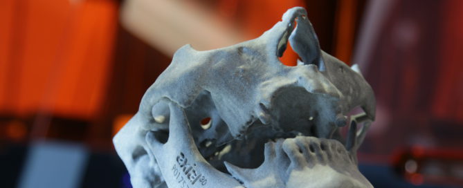
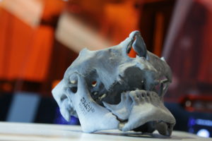
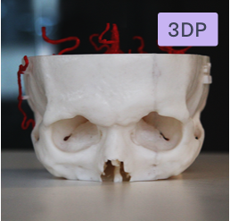
A 3D printing lab at the institute can produce replicas of organs, regulatory systems and other body parts using X-rays and other radiological images of patients about to undergo surgery. The malleable models feel rubbery, real. They replicate all the nooks and crannies of an individual patient’s anatomy. Specialists scheduled to operate can use the simulator and models to get a feel for potential trouble spots; say an unusual bend, bulge or narrowing in an artery, or a lesion inside an organ.

“We figure out where the hurdles are in the model first,” Levy said, “and it’s usually smooth sailing with the patient.”

The 3D lab contains two printers. The most sophisticated, a Stratasys Ltd. 3D printer-maker, can produce objects in color. “At the GVI, we do training for physicians and medical device manufacturers all over the world using this technology,” said Levy, also president of the UB neurosurgery physicians’ group. “Now, potentially, we could use similar technology to educate laypeople.”
The attorneys jumped at the chance for Gates Vascular and UB to test the idea that two models of their client could be turned into evidence for their court case. Normally in substantial personal injury cases that appear headed for trial, lawyers pay medical illustrators up to $10,000 to gather patient records and images, interview treating physicians and create one-dimensional drawings that show specific injuries. Generic body part models often are used for comparison. Attorneys then use black-and-white X-rays, MRIs and CT scans, as well as written medical reports, to bolster their case, said Fromen, a 32-year legal veteran.
Attea called the new approach “the CSI effect.”

Focus groups

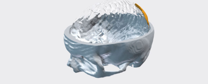
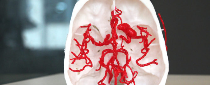
Fromen and Attea wanted to show the models to focus groups to see how jurors might respond. One model depicted injuries to the arterial system inside the neck and skull. The second showed damage to the upper spinal column, where a ligament was torn from the spinal column, damaging several discs. The first models were produced in an off-white shade, which at times confused
some of the first of seven focus groups to see them. Using input from those groups, UB mechanical engineers produced colored models in which key body components better stood out. Spinal vertebrae remained white. Spinal discs, normally yellowish, were turned blue. A splash of red showed blood in the spinal column caused when the ligament tore loose.
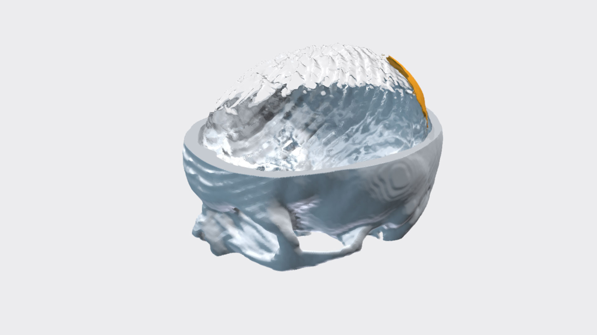
The model of the arterial system was produced in pale red. Two small aneurysms behind the eyes remained white. The one Levy discovered behind the left eye was larger than the one behind the right. Had he been called to testify, he would have explained that they would have been the same size had they formed congenitally before the crash, and that a traumatic collision best accounted for them.

“Prior to this, we never had the ability to show jurors what an aneurysm looks like,” Fromen said. “This is my client’s actual anatomy down to thousands of sub-millimeters.”

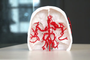
Another idea resulted in cutting the rectangular model of the spinal column in half and adding a hinge, so injuries could be seen from the inside. “The doctor can get off the witness stand,” Fromen said, “show the model to the jury and say, ‘This is her spinal cord, and these are the damaged discs impinging on the spinal cord. That’s dangerous, because it could result in paralysis.’ It’s amazing how small someone’s spinal cord actually is and how the external and internal carotid arteries go to the neck and supply blood into the brain, and you can see it in these models.”

The future

Fromen and Attea settled the case for $3.5 million during a mediation conference on the eve of trial. It was the first time the defendant’s attorney and a representative from the insurance company involved in the case saw the 3D models. The computer-generated replicas, and the prospect that a leading surgeon would testify that injuries depicted were caused by the truck- pedestrian crash, were key to the settlement, lawyers for the plaintiff said.
The Lancaster crash victim and her husband, an engineer, were fascinated with the models and grateful they helped reach an acceptable settlement. Still, their lawyers said, it would have been better for them had a stranger made better choices while driving a slippery road three years ago.

“My client used to walk a lot,” Fromen said. “Now she can’t walk for extended periods of time or on uneven surfaces for a couple reasons. Number one is because the problems she has with her right leg, but her biggest fear is falling. If she falls, she’s got plates and screws in her neck. She could be paralyzed.”

Her experience after the crash may help others in the future who befall a similar fate, her lawyers said. UB and Gates can now produce the 3D models economically, Levy said, “but if somebody wanted to create these models for use in a court case, and turn it into a business, 3D printers are pretty expensive.”

Tags

Fromen and Attea believe someone will make such an investment, and likely become the go-to company to bolster medical-related legal cases. In fact, opportunities like this were what UB planners hoped for in 2012, when the university opened the Clinical and Translational Research Center on the floors above the Gates Vascular Institute.
Story originally appeared in Buffalo news 
Up next: Discover the differences between 2D images and 3D printing in the courtroom
2021-05-21T12:37:27+00:00February 23rd, 2021|
Go to Top